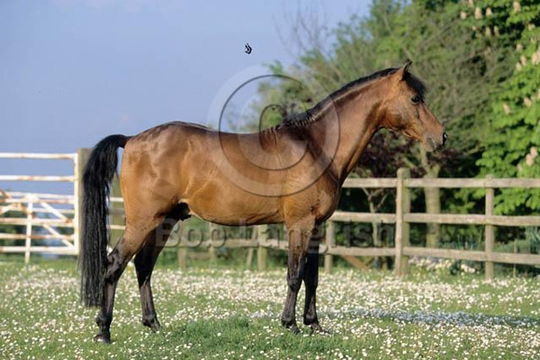
The histological structure of the lymphatic follicles and their distributions pattern throughout the cecum in five healthy mature miniature horses were studied.
Article in Anantomia Histologia Embryologia
Maryam Rezaian , University of Tehran
A. Saheb Jamei
F. Ebrahimpoor
Abstract
The cecum is a great cul-de sac intercalated between the small intestine and the colon. It has a remarkable size in the equine and digestion of more than 30% of total cellulose and production of volatile fatty acids by anaerobic metabolism of cellulose by its micro flora were made on it. It has also a substantial number of lymphatic nodules, which in horses are located near the apex. Topographically, cecum is situated chiefly to the right of the median plane and occupies around the whole volume of there in the abdominal cavity.
The histological structure of the lymphatic follicles and their distributions pattern throughout the cecum in five healthy mature miniature horses (an special species of horses lives only in north of Iran) were studied (Fig. 1a). Each cecum was divided into three parts, namely, the base, body and apex, and samples were taken from each part immediately after slaughter and fixed with 10% buffered formalin. Routine histological laboratory techniques were made and 6 m ?sections were cut and stained with haematoxylin and eosin staining and studied under light microscope.
Lymphatic nodules were mostly found in the submucosa, but they disrupted the muscularis mucosa and merged into the lamina propria (Fig. 1c). The thickness of the mucosa and submucosa at these sites was smaller, and the epithelium that covered the mucosa was here modified into simple squamous or low cuboidal epithelia with dark and flattened nuclei (Figs 1b,d). The large and small lymphatic nodules were extensively accumulated in the apex, forming a nearly continuous layer in lamina propria and submucosa. However, they were less prominent at the base and least in the body of the cecum.