In Caspian miniature horses images obtained from the left side were of poorer quality than those obtained from the right.
Two dimensional (2-DE) and M-mode echocardiography were performed on 33 unsedated, healthy Caspian miniature and 10 thoroughbred horses.
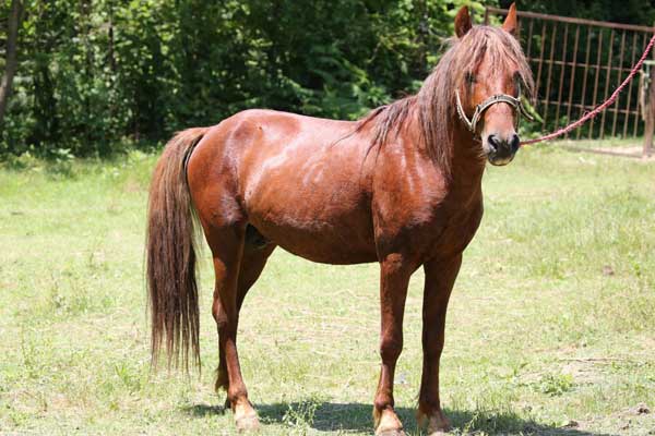
The groups comprised 26 adult, 7 young Caspian and 10 adult thoroughbred horses. Animals stood during examinations performed with a 3.5 MHz Mechanical sector transducer using various echocardiographic views. Images were recorded from the right and left sides of the thorax. 2-DE images were used to guide the placement of a cursor to obtain accurate M-mode recordings.
The recommendations of the American Society of echocardiogrphy were followed for all M-mode measurements. The leading edge method was used for all M-mode measurements. Right and left ventricular diameters, ventricular septa thickness (IVS) and ventricular free walls thickness were measured in chordal level with the axial beam prependicular to interventricular septum in end systole and end diastole Echocardiographic parameters of mitral and aortic valves and
left atrium were calculated end systole and diastole. Using a 3.5 MHz probe, the quality of recording were adequate for quantitative and qualitative examinations.
In Caspian miniature horses images obtained from the left side were of poorer quality than those obtained from the right. Summary statistics for each dimensions were determined including the range, mean, standard deviation, standard errors of mean and coefficient of variance.
Significant differences between mean value of cardiac dimensions in adult Caspian miniature and thoroughbred horses were determined using The student-T test (P
Keywords: 2-DE, Caspian miniature horse, echocardiography, M-mode, Standard

