The Caspian miniature horse is one of the rare small breeds in the north of Iran. In the present study, the position of the heart and its valves were determined topographically in 4 miniature horses. We found that Caspian miniature horses have general similarities, with certain topographical variability, with other horses.
M.R. Paryani, H. Gilanpour
Department of Basic Sciences, Faculty of Veterinary Medicine, University of Tehran, Iran
Address for correspondence: M.R. Paryani, Department of Basic Sciences, Faculty of Veterinary Medicine, Islamic Azad University, Karaj Branch, Karaj, Iran, tel: +98 261 441 81 43, fax: +98 261 441 81 56, e-mail: mrparyani@kiau.ac.ir
[Received 29 July 2008; Accepted 19 November 2008]
Folia Morphol.
Vol. 68, No. 1, pp. 36–39
Copyright © 2009 Via Medica
ISSN 0015–5659
www.fm.viamedica.pl
INTRODUCTION
The Caspian miniature horse, an indigenous breed of horse from the north of Iran, is believed to be the ancestor of many current breeds [4]. It is suggested that the miniature horse is possibly the offspring
of a natural hybridization between E. caballus and E. przewalski [6, 7]. The postural statuses of
miniature horses are similar to those of non-miniature horses. Apart from height, however, minor anatomical differences have been reported between miniature horse and Equus caballus [3].
The location of the heart and the areas of auscultation relative to the underlying valves are very important in detecting the heart sound, as well as in echocardiography [2, 9, 10]. However, little is
known about the topographical position of the heart and areas of auscultation of Caspian miniature horses.
The purpose of the present study was to determine the topographical location of the heart and its valves in the Caspian miniature horse by anatomical dissection.
MATERIAL AND METHODS
Four miniature horses were obtained from the Caspian Miniature Horse Centre, Khojeer, Iran in 2005. The animals were anesthetized, and the carotid artery was exposed and canulated for exsanguination. Three out of four horses were fixed by injection of embalming fluid into the carotid artery.
A solution was made with the following substances: 200 g Potassium acetate and 100 g Thimole dissolved in 500 mL of H2O; 1000 mL Formalin (40%); 500 mL Phenol (liquid); 500 mL Dettol; 1000 mL Glycerine. Ethyl alcohol 95% was added to the solution to obtain 20 L.
The embalmed horses were kept at 3°C for 30 days. The fourth horse was dissected fresh after
exsanguination. Dissection was carried out in the standing position. The skin was removed and the
thorax was dissected layer by layer on both sides. After removing the muscles, the position of the lung and cardiac notch were determined with regard to the ribs.
To determine the topographical position of the heart and its valves, the 2nd, 3rd, 4th, 6th, 7th, 9th, and
11th ribs were removed by cutting below the vertebral end and above the costochondral junction parallel
to the line of pleural reflection.
To expose the heart, the lungs were also removed. The pulmonary, aortic, and bicuspid valves were
demonstrated on the left side by partly removing the wall of the pulmonary artery, the aorta, and the
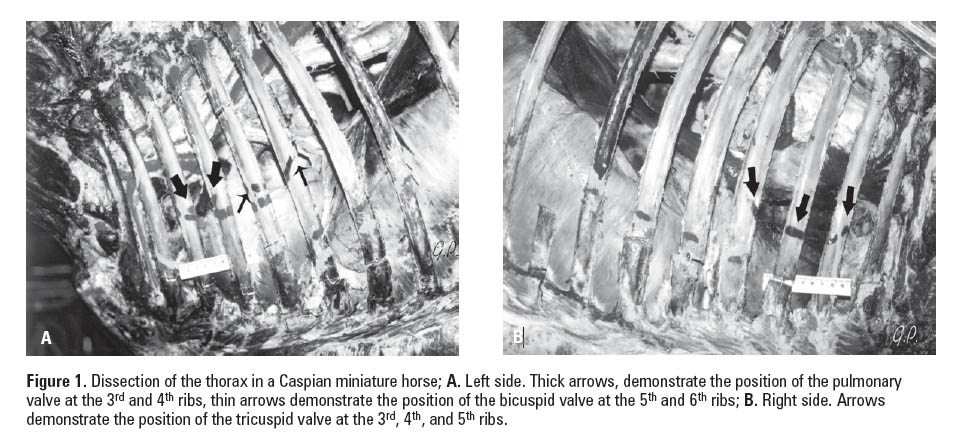
left atrium. The tricuspid valves were exposed on the right side when the wall of the right atrium was partly removed. The ribs were placed in their original position. The base of the heart, the apex, and
location of the valves were marked on the ribs and were clearly measured according to the ribs and intercostal spaces (Fig.1A, B).
All stages of the dissection were photographed and the procedure was recorded.
RESULTS
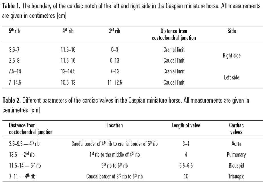
Cardiac notch
The cardiac notch on the right side at its maximum width was extended from the 3rd to 5th rib. On
the left side, the maximum width was mainly extended from the 3rd to 5th rib, but in one case the
cranial limit was at the 2nd rib and in another case the caudal limit was at the 6th rib. The distance of
the cranial and the caudal limits on the left and the right side from the costochondral junction is demonstrated in Table 1.
Base of the heart
The base of the heart was extended from the 2nd intercostal to the 5th intercostal spaces on the right
side. On the left side, it was extended from the 2nd to the 5th intercostal spaces in one case and from
the 2nd to the 6th intercostal spaces in two other cases. The distance from the costochondral junction at
the 6th rib varied from 13 to 17 cm.
Apex of the heart
The apex of the heart was situated at the level of the 5th costochondral junction. In two cases it was
extended briefly to the 4th intercostal cartilage space. Different parameters and position of pulmonary,
aortic, bicuspid, and tricuspid valves were as follows and are demonstrated in Table 2.
Pulmonary valve
The pulmonary valve was exposed on the left side of the thoracic cavity. The length of the valve measured 4 cm and extended from the 3rd to the cranial half of the 4th rib. It was situated 9.5 to 13.5 cm
from the costochondral junction at the 3rd intercostal space (Fig. 1A).
Aortic valve
The aortic valve was exposed on the left side, after removing the pulmonary artery and cutting a window in the aortic valve below the brachiocephalic trunk. In two cases, it extended from the
caudal border of the 4th rib to the cranial border of the 5th rib (4th intercostal space). In one case, the
cranial limit was at the cranial border of the 4th rib. The length of the valve was 3 to 4 cm and was
situated 9.5 to 12.5 cm from the 4th costochondral junction.
Bicuspid valve
The bicuspid valve was exposed on the left side. It extended from the 5th rib to the 6th rib. The length
varied from 5.5 to 6.5 cm. The distance from the costochondral junction varied from 11.5 to 14 cm
at the 5th intercostal space (Fig.1A).
Tricuspid valve
The tricuspid valve was exposed on the right side. It extended from the caudal border of the 3rd rib to
the 5th rib. The length was 10 cm. The distance from the costochondral junction varied from 7 to 11 cm
at the 4th rib (Fig. 1B).
DISCUSSION
The cardiac notch. The area of the heart which was not covered by the lung was mainly extended on both sides from the 3rd to the 5th rib at its maximum width, which is similar to what has been described for the horse [1, 5, 8]. However, unlike that of the horse, there was variation from the 2nd to the 6th rib on the left side. The cranial and caudal boundary of the cardiac notch is shown in Table 1.
The results indicates that the cranial border of the notch on the right side begins at the costochondral
junction of the 3rd rib or a little higher (0 to 3 cm), and the maximum height of the notch 11.5 to 16 cm from the costochondral junction is at the 4th rib. The caudal border extends steeply and ventrally to
reach the caudal border of the 5th rib at a level of 2.5 to 8 cm from the costochondral junction.
The cranial border of the notch on the left side begins at 11 to 12.5 cm from the costochondral junction
of the 3rd rib, and the maximum height of the notch is at the 4th rib, 10.5 to 14.5 cm from the costochondral junction. The caudal border of the notch extends steeply to reach the 5th rib at 7 to 14.5 cm from costochondral junction.
Knowing the position of the cardiac notch might be important from a topographical point of view as there seems to be lack of information on this parameter on other breeds of horse. The position of the base of the heart was from the 2nd to the 5th intercostal spaces on the right side but it may reach the 6th intercostal space on the left side in the Caspian miniature horse.
The report on the horse shows that the base of the heart extends from the 2nd intercostal space or the
3rd rib to the 6th or sometimes the 7th rib [5]. This seems slightly greater than the Caspian miniature horse.
The apex of the heart in the horse is at the level of the 5th costochondral junction [5]. However, it has
been reported that it might be situated in the 6th costal cartilage [3]. Our result shows that the apex of the heart in the Caspian miniature horse is at the 5th costochondral junction, like that of the horse, but it might be situated at the 4th costochondral junction, which seems to be slightly more cranial than that of the horse.
The pulmonary valve in the Caspian miniature horse was extended from the 3rd to the cranial half of the 4th rib, which is slightly more caudal than that of the horse, which extends from the 3rd rib to the 3rd intercostal space [1]. The position of the aortic valve, similar to that of the horse, was medial to the pulmonary artery [1]. The cranial limit of the aortic valve may begin from the cranial border of the 4th rib and extends to the cranial border of the 5th rib, which shows the location is at 5th intercostal space; this seems to be different from that of the horse, which is reported to be situated from the 5th rib to the 5th intercostal space [5].
The bicuspid valve is similar to what has been reported in the horse [1, 5].
Tricuspid valve. In the Caspian miniature horse, the tricuspid valve extended from the 3rd rib to the 5th rib, which seems to be different than that of the horse, in which it is mainly situated at the 6th rib [1, 5].
ACKNOWLEDGEMENTS
This study was funded by the Ministry of Science, Research, and Technology and the research was arried
out at the anatomy laboratory of the Veterinary Faculty of the University of Tehran.
The horses were provided by the Caspian miniature horse breeding centre, Khojeer, Iran. We are thankful to Dr. Dordary for these arrangements. We are also thankful to Mr. Sabouri and Mr. Chavoshi
for their technical assistance.
REFERENCES
1. Budras KD, Sack WO, Röck S (2003) Anatomy of the horse. 4th Ed. Schlûtersche GmbH & Co.KG, Verlag und Druckere, p. 56.
2. Detweiler DK (1958) Examination of the equine heart. Univ. Pennsylvania Bull. Vet. Extension Quarterly No. 152.
3. Dyce KM, Sack WO, Wensing CJG (2002) Textbook of veterinary anatomy. 3rd Ed. Sunders Co. Philadelphia, pp. 219–221.
4. Firoz L (1971) Osteological and histological implication of the Caspian pony to early domestication in Iran. Proc. 3rd Int. Congr. Agricultural Museum, Budapest, pp. 1–5.
5. Firous L (1998) The original ancestors of the Turkoman, Caspian horses. Proc. 1st Int. Conference on Turkoman Horse, Ashgabad, Turkmenistan, May.
6. Getty R (1975) Sisson and Grossman’s anatomy of the domestic animals. 5th Ed. W.B. Saunders Co. Philadelphia, pp. 554–570.
7. Glazier DB (1987) Clinical aspects of equine cardiology. Practice, 9: 98–103.
8. Hatami-Monazah H, Pandit RV (1979) A cytogenic study of the Caspian pony. J Reprod Fert, 57: 331–333.
9. O’Callaghan MW (1993) Comparison of echocardiographic and autopsy measurements of cardiac dimensions in the horse. Equine Vet J, 15: 38–68.
10. Patteson MW, Cripps PJ (1993) A survey of cardiac auscultator findings in horses. Equine Vet J, 25: 409–415